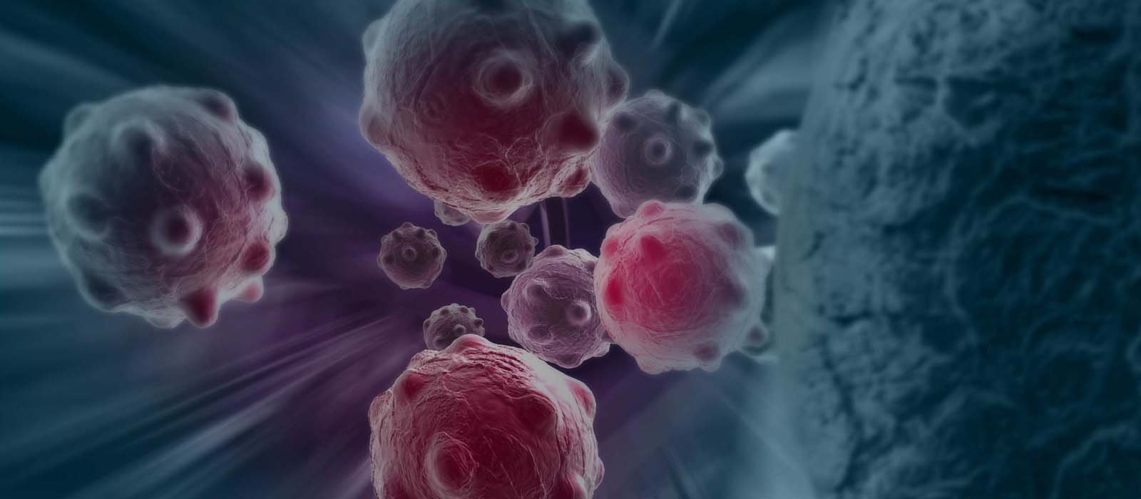
Giant Cell Tumor is a benign (non-cancerous) aggressive tumor of bone. It is most common benign bone tumor. its commonly seen in the age group of 20 to 40 years. It is seen at the end of long bones. I.e, just above or below the joint line. Its commonest location is lower end of femur bone or upper end of tibia, i.e, the bone just above or below the knee joint. However, it can affect any bone and any age group of patient.
Exact cause of Giant Cell Tumor is unknown. However, it has been linked to genetic mutations and also calcium metabolism disturbances.
It is diagnosed with the help of Xray, MRI and a needle biopsy – histopathology.
GCT being a noncancerous tumor, very rarely spreads to the other parts. Hence, its staging is not done. However, in about 2% of patients, it can spread to lungs or neighboring lymph nodes. It’s also known to be multicentric i.e, in more than 1 bones.
On the bases of its xray picture, it is divided in 3 types.
Stage 1: The tumor is restricted within the boundaries of the bone.
Stage 2: Tumor is expanding the bone deforming its contour but hasn’t breached the wall of the bone.
Stage 3: Tumor has destroyed one / more walls of bone and has presented with a soft tissue mass outside the bone.
As GCT is a benign tumor, the goal of treatment would be to conserve the neighboring joint and retain patient’s normalcy as much as possible. The treatment of Giant Cell Tumor consists of a surgery.
A special type of surgery is offered to the patients of GCT which is known as “Radical Intra-lesional curettage”.
A large window is made in the wall of the bone to expose all the nooks and corners of the disease cavity. All the tumor tissue is removed in pieces with help of variety of instruments known as scoops. A surgeon will be able to remove what is visible to him. However, if the procedure is concluded at this point, the microscopic tumor (which is not visible naked eye) will be left behind. This will lead to recurrence of the tumor. Conventionally, the recurrence rates reported in medical literature are in range of 40 to 60%!
“Radical Intra-lesional curettage”. surgery tackles this microscopic disease. Some adjutants are used to kill the microscopic disease. mechanical agents like A high speed burr, thermal agents like laser / electrocautery and chemical agents like phenol are used to kill the residual tumor cells. With this kind of aggressive removal of even microscopic tumor cells, the recurrence rates have reduced to less than 5%!
Once the curettage is done, the defect or cavity is filled with either Bone grafts (harvested from patient’s own body) or bone cement. In some patients, plate fixation id also done to augment the strength to the construct.
In very large giant cell tumors, sometimes the curettage method is not possible or unsafe. In this kind of situation, complete resection of the tumor is done followed by reconstruction with joint replacement or other methods.
Once the treatment is over, patient needs to follow up on regular bases irrespective of any problems for next 5 years. It is only then, that the patient is declared free of tumor!
| 1 | For first 2 years | 3 monthly |
|---|---|---|
| 2 | For next 3 years | yearly |
| 3 | For next 5 years | sos |


